
Subdural Hematoma (SDH) YouTube
Epidural hematomas (EDHs) and subdural hematomas (SDHs), or so-called extra-axial bleedings, are common clinical entities after a traumatic brain injury (TBI), and both are often present in the same subject [ 1 ].
Difference between Extradural and subdural Hematoma MEDizzy
For more video tutorials, high-yield revision Qbanks and support from our online Community, Sign Up at https://www.surgicalteaching.com. Use Coupon code STYo.

Clinics in neurology and neurosurgeryextradural and subdural haematoma Davis et al. 44 (16
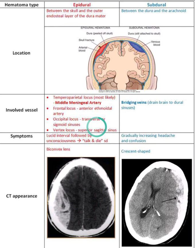
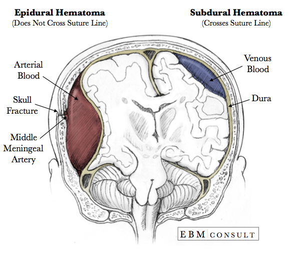
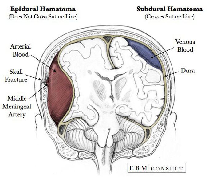
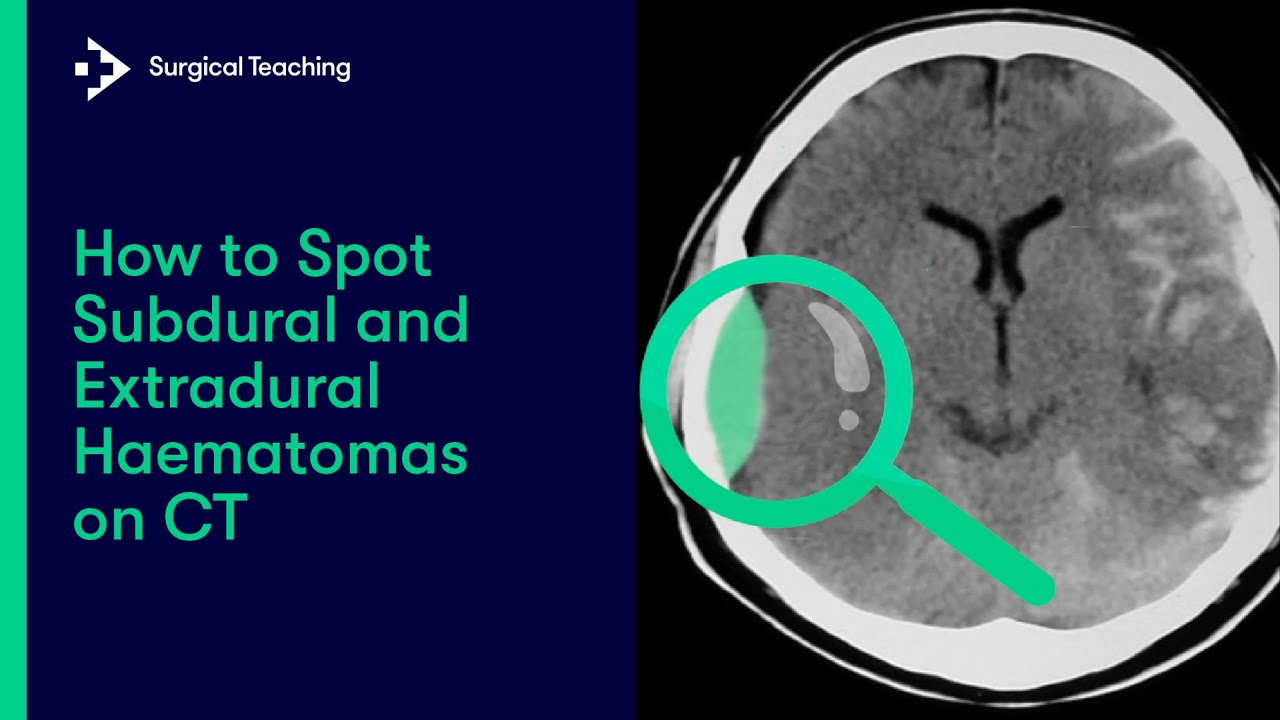
Subdural hematomas are interposed between the dura and arachnoid. Typically crescent-shaped, they are usually more extensive than extradural hematomas. In contrast to extradural hemorrhage, subdural hemorrhage is not limited by sutures but is limited by dural reflections, such as the falx cerebri, tentorium, and falx cerebelli.
Acute CT Brain Extraaxial haemorrhage
A venous epidural hematoma occurs when there is a skull fracture, and the venous bleeding from the skull fracture fills the epidural space. Venous epidural hematomas are common in pediatric patients. Subdural Hematoma Subdural hemorrhage occurs when blood enters the subdural space which is anatomically the arachnoid space.
Anatomy Epidural vs Subdural Hematoma Image
Duret hemorrhages are associated with descending transtentorial herniation, which can occur due to various underlying causes. Herniation syndromes manifest as a result of increased intracranial pressure, leading to shifts in intracranial compartments. The etiology of Duret hemorrhages include 8: epidural hemorrhage. subdural hemorrhage.
Epidural Hematoma vs. Subdural Hematoma MEDizzy
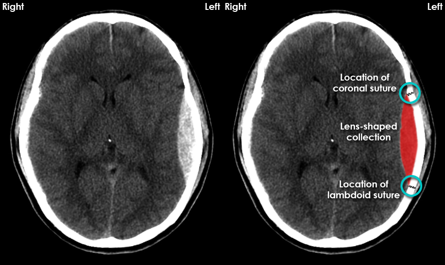
An extradural haematoma (also termed an epidural haematoma) is an extra-axial bleed occurring between the dura and skull bone. Around 2% of all head injuries presenting to the emergency department are extradural haematomas (EDHs), with associated significant morbidity and mortality, especially with advancing age.

subdural hematoma vs epidural hematoma XRAY Google Search Neuro Pinterest Grey, Old
A subdural hematoma is a type of brain bleed. Blood leaks out of a blood vessel into the space below the outermost membrane of the brain -- the dura mater. What is a subdural hematoma? A subdural hematoma is a type of bleed inside your head. It's a type of bleed that occurs within your skull but outside the actual brain tissue.

Pin on Computed Tomography
Extradural haematoma (EDH) is defined as an acute haemorrhage between the dura mater and the inner surface of the skull. An EDH can cause compression of local brain structures and a rise in intracranial pressure. If intracranial pressure continues to rise, cerebellar herniation may occur leading to brainstem death.

Pin on Medicina
A subdural hematoma forms because of an accumulation of blood under the dura mater, one of the protective layers to the brain tissue under the calvarium. The understanding of subdural hematoma relies on the knowledge of neuroanatomical sheets covering the brain. The brain is the central repository of delicate neural tissue. This network of neurons and neuronal connective tissue is prone to.

Download Subdural vs Epidural Hematoma/Hemorrhage [CT Scan Findings] Watch online
Extradural Hematoma vs. Subdural Hematoma. A hematoma is a medical term for a blood clot, formed when bleeding occurs in an organ or cavity. Extradural hematoma or epidural hematoma (EDH) is a clot that forms on the external side of the brain's protective layer of tissue (dura mater), whereas acute subdural hematoma (ASDH) appears within the first few days after head injury, on the interior.

SUBDURAL VS EPIDURAL HEMATOMA RADIOLOGY Wroc?awski Informator Wroc?aw, Wroclaw
Introduction A subdural haematoma, also known as a subdural haemorrhage, is defined as a collection of blood between the dura mater and arachnoid mater of the brain. 1 A subdural haematoma (SDH) can be acute (< 3 days), subacute (3-21 days) or chronic (>21 days).

EPIDURAL & SUBDURAL HEMATOMA Medical Facts, Medical Science, Computer Science, Icu Nursing
1 Images Snapshot A 76-year-old man presents to the emergency department with increasing somnolence and lethargy. His symptoms developed on the day of admission. His symptoms are associated with a headache with mild nausea but no vomiting. He tripped over a carpet and hit his head on the floor.
Extradural vs Subdural Haematomas How do we Diagnose them on CT? YouTube
SDH is a collection of blood between the dural and arachnoid coverings of the brain. As the volume of the hematoma increases, brain parenchyma is compressed and displaced, and the intracranial pressure may rise and cause herniation. While the presence of SDH can be inferred by neurologic decline and mechanism of traumatic injury, the diagnosis.

CNS Trauma Pathoma
Both extradural and acute subdural haematoma are serious conditions that can cause compression of the underlying brain, and usually are a result of significant head trauma, which may be associated with a brief episode of loss of consciousness. Causes Extradural haematoma (usually from a torn artery in the skull)… Head trauma. Fracture of skull.

Subdural Hematoma Vs Epidural Hematoma slidesharetrick
A subdural hematoma occurs when a blood vessel near the surface of the brain bursts. Blood builds up between the brain and the brain's tough outer lining. The condition is also called a subdural hemorrhage. In a subdural hematoma, blood collects immediately beneath the dura mater. The dura mater is the outermost layer of the meninges.

Head injury in adults Induction RCEMLearning India
Overview An intracranial hematoma is a collection of blood within the skull. It's usually caused by a blood vessel that bursts in the brain. It may also be caused by trauma such as a car accident or fall. The blood may collect in the brain tissue or underneath the skull, pressing on the brain.